What is patellar instability?
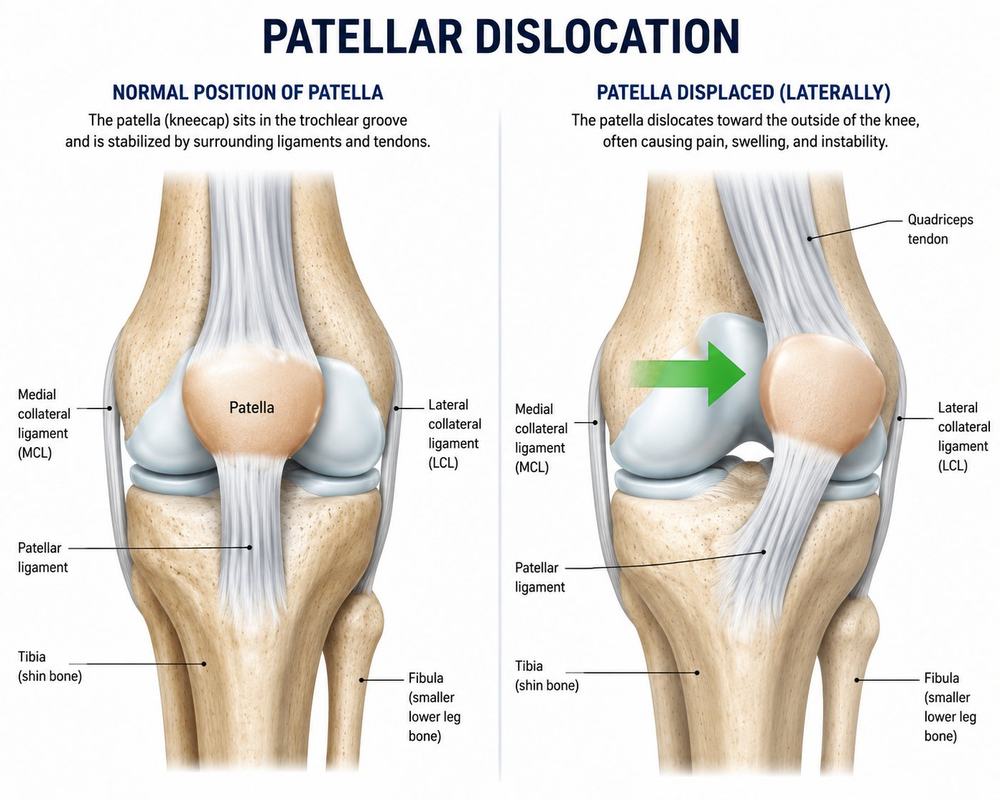
Patellar instability occurs when the kneecap slips partially (subluxation) or fully (dislocation) out of its groove on the femur. It's most common in adolescents and young athletes, and usually reflects a combination of anatomic factors — a shallow trochlear groove, ligament laxity, patellar height, or overall alignment — rather than a single cause. The medial patellofemoral ligament (MPFL), the main checkrein against the kneecap shifting outward, is torn in nearly every dislocation.
Typical symptoms: a visible or felt shift of the kneecap, immediate swelling after a dislocation, a sense the kneecap might give way, and pain with stairs or kneeling.
How is it evaluated?
Exam assesses kneecap tracking and apprehension with lateral movement. MRI evaluates the MPFL, the cartilage surfaces (which can be injured during a dislocation), and the underlying anatomic risk factors such as groove depth and patellar height — the factors that drive both recurrence risk and the choice of procedure.
Treatment options
A first-time dislocation is often treated without surgery: bracing and physical therapy focused on strengthening and alignment. Recurrent instability — especially with underlying anatomic risk factors — is treated surgically:
- MPFL reconstruction rebuilds the torn checkrein ligament with a graft, restoring the primary restraint against dislocation.
- Tibial tubercle osteotomy (TTO) repositions the bony attachment of the patellar tendon when malalignment contributes, improving tracking and unloading the cartilage — often combined with MPFL reconstruction for a comprehensive correction.
- Cartilage damage sustained during dislocations can be addressed at the same surgery.
Recovery & return to sport
After surgery: bracing and protected weight-bearing for roughly the first 6 weeks, progressive strengthening and motion over months 2–4, sport-specific training months 4–8, and return to cutting and pivoting sports guided by strength testing rather than the calendar.
Pain control after surgery
Frequently asked questions
Will my kneecap dislocate again?
Recurrence risk is higher after a first dislocation, especially in younger patients and those with anatomic risk factors — which is why targeted rehab, or surgery for recurrent instability, is recommended.
Do I need surgery after one dislocation?
Not always. Many first-time dislocations are treated non-surgically with bracing and therapy; surgery is considered for recurrent instability, loose cartilage fragments, or high-risk anatomy.
What does the MPFL do?
The medial patellofemoral ligament is the primary soft-tissue restraint keeping the kneecap from shifting outward — it tears in nearly every dislocation, and reconstructing it restores that checkrein.
When is bone surgery (TTO) added?
When imaging shows the tendon attachment sits too far lateral or other bony malalignment, moving the tibial tubercle corrects the direction of pull — soft tissue alone can't fix a bony problem.